|
|
Atrial Septal Defect
ASD
General Considerations
- Atrial septal defects (ASD) represent 10-15% of congenital cardiac abnormalities
- Sinus venosus type of ASD accounts for 10% of ASDs and only 1% of all congenital heart lesions
Four Major Types of ASD |
- Most common is ostium secundum (60%) located at fossa ovalis
|
High association with prolapse of the mitral valve |
- Ostium primum type is usually part of an endocardial cushion defect
|
It tends to act like a VSD physiologically |
- Sinus venosus type lies high in the interatrial septum
|
90% association of anomalous drainage of R upper pulmonary vein with SVC or Right atrium |
- Most rare type is posteroinferior ASD
|
Associated with absence of the coronary sinus and a left SVC emptying into LA |
- 2:1 ratio of females to males
- The most frequent congenital heart lesion initially diagnosed as an adult
- Sinus venosus type occurs high in the inter-atrial septum, in contiguity with the superior vena cava
- Invariably associated with anomalous pulmonary venous drainage of the right upper pulmonary vein into the SVC (partial anomalous pulmonary venous return-PAPVR)
Clinical Findings
- Usually asymptomatic in infancy
- Cardiac murmur
- Easy fatigability
- Dyspnea
- If uncorrected, arrhythmias, congestive heart failure and eventually pulmonary hypertension in adulthood
Imaging Findings
- Chest radiographs may be normal or have an enlarged main pulmonary artery and increased pulmonary vasculature
- Echocardiography will show ASD and most pulmonary vein connections
- MRI will show ASD and outline venous anatomy
- Septal defect high in the inter-atrial septum contiguous with superior vena cava
- Posterior to location of fossa ovalis
Differential Diagnosis
- Other forms of atrial septal defect
- Partial anomalous pulmonary venous return without ASD
Treatment
- Sinus venosus ASDs do not close spontaneously
- Surgical repair in the first two decades of life
- A patch of either synthetic material or pericardium redirects blood flow from the right superior pulmonary vein into the left atrium.
Complications
- Congestive heart failure
- Pulmonary arterial hypertension
Prognosis
- Excellent prognosis if treated


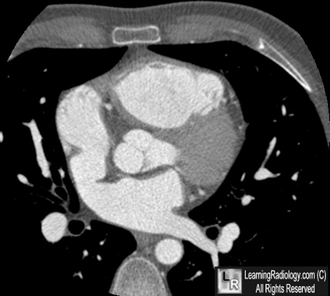
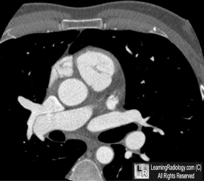
Sinus Venosus Atrial Septal Defect with Partial Anomalous Pulmonary Venous Return. Top: There is a large atrial septal defect (red arrow) connecting the left atrium (LA) and right atrium (RA) at the level of the superior vena cava. Bottom: Right pulmonary veins drain directly into the superior vena cava (S) instead of the normal return to the left atrium.
For these same photos without the arrows, click here and here
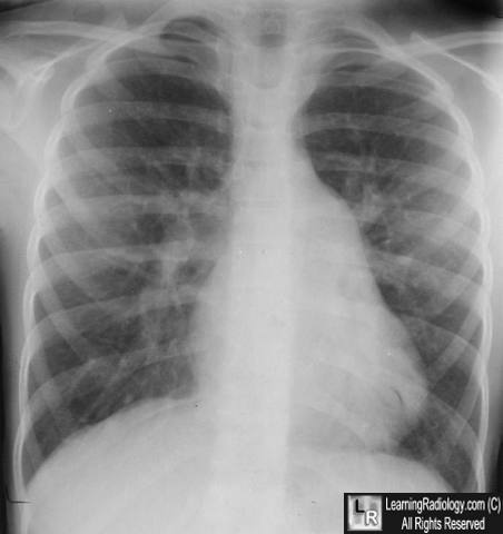
Atrial Septal Defect. There is a large atrial main pulmonary artery because of the increased shunt from left to right. The pulmonary vasculature is prominent throughout the lungs due to increased flow.
For more information, click on the link if you see this icon 
Atrial Septal Defect, Sinus Venosus. eMedicine. GM Satou
|
|
|
